Dental X-rays
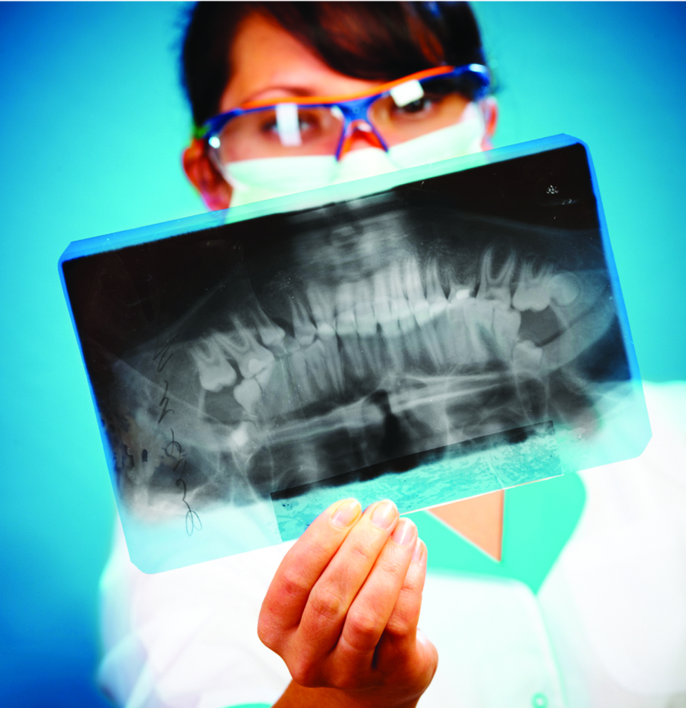
Dental x-rays are images of your teeth so that Dr. Dennis can evaluate your oral health.
X-rays are used with low levels of radiation to capture images of the interior of your teeth and gums.
Why it's important to have a dental x-Ray?
Dental x-rays are used to diagnose issues that are nearly invisible to the naked eye; things like cavities, tooth decay, and impacted teeth.
With the Help of X-rays We Can See:
- Decay, between teeth or underneath a filling
- Bone loss associated with gum disease
- Abscesses, infections at the root of the tooth
- Abscesses, infections between the tooth and gum
- Identify tumors within oral tissue
- Changes in the root canal
- Identify cavities and view tooth roots
- Assess the health of bone around the teeth
- Identify periodontal disease
- Evaluate the status of developing teeth
Without an x-ray, many of these problems may not be diagnosed.

With x-rays we are better equipped to prepare tooth implants, dentures, braces, and other treatments.
Two Main Types of Dental X-rays:
Intraoral, meaning the X-ray film is inside the mouth, the most common type of dental X-ray taken.
Extraoral, meaning the X-ray film is outside the mouth.
Three Types of Diagnostic X-rays:
Common Questions About X-Rays
Dental X-rays are considered safe for the vast majority of healthy people. Dental professionals abide by strict safety guidelines to minimize a patient’s exposure to radiation. If you have a pre-existing medical condition or are undergoing cancer treatment, tell Dr Dennis before your exam.
Dental X-rays are one of the smallest radiation-dose studies performed in modern medicine. A routine exam with four Bite-Wings transmits about 0.005 mSv, which is very similar to the amount of radiation a person would be exposed to during a plane flight of one to two hours.
According to the ADA, research has affirmed that dental X-rays are safe for pregnant women and their fetuses. Other research has also shown that it’s safe for pregnant women to undergo dental treatment with local anesthetics. That said, you should always tell your local dentist if you are pregnant or breastfeeding before you get an exam.
The need for x-rays is based on age, risk, and signs of disease or oral issues. Healthy patients with little cavity risk may have x-rays performed every year or two, while high risk patients may receive them more often (possibly every 6 months). The ADA recommends that dentists and patients discuss the need for x-rays together.
Dental X-rays are safe during pregnancy. In fact, it’s very important to have dental X-rays done while pregnant. X-rays are an effective diagnostic tool that helps detect tooth damage and disease not otherwise visible during a regular dental exam. If there’s an unseen oral health issue, it’s crucial to diagnose it before it affects both the mother and the baby. Because the mouth is the gateway to the body, an infection in the mouth can spread to other parts of the body—including the baby. The radiation produced during an X-ray won’t harm the mother or the baby because the technician routinely places a lead apron and thyroid collar to protect the abdomen and thyroid.
The cost of a dental X-ray can vary from $15 to $200 dollars, depending on the type of X-ray and your city or state. Most dental insurance plans will cover all or most of this expense, as long as you haven’t exceeded your limits for a given year.
